The photo that convinced you
Most patients can name, at least roughly, the image that started their treatment journey. A before-and-after pair on a clinic website, a comparison shared on social media, a screenshot sent by a friend. Something in that image made a previously abstract possibility feel concrete. The gap between the two photographs gave the treatment a shape.
There is nothing inherently wrong with that. Clinical photography is a legitimate communication tool, and a well-constructed gallery can convey information that words cannot. The problem is that before-and-after photographs are also among the most easily manipulated visual formats in existence — not necessarily through fraud, but through the accumulation of small, technically defensible choices that collectively produce an unreliable impression. Lighting change here, slight angle tilt there, a different time of day, a different skincare routine in the “after” morning — and the result looks more dramatic than the treatment alone could explain.
Understanding how this manipulation works is not about distrust. It is about becoming the kind of patient who can evaluate a gallery accurately, ask the right questions, and set expectations that the treatment can actually meet.
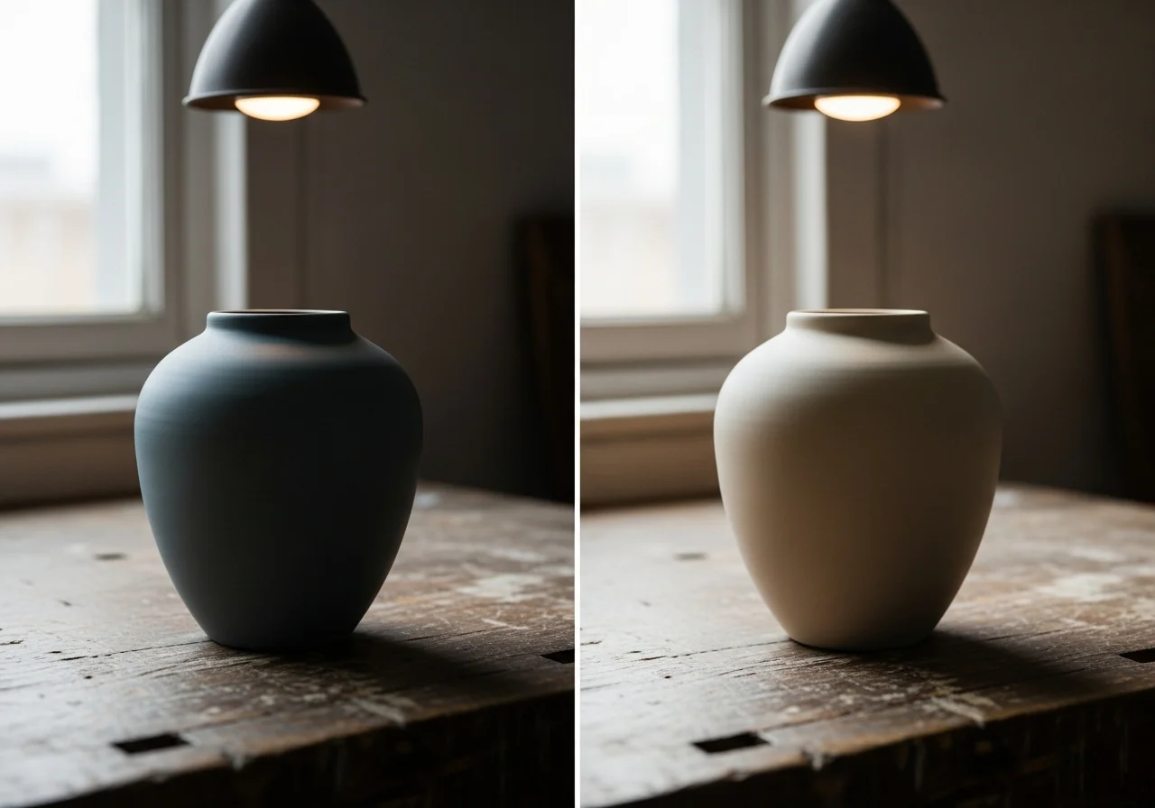
How does lighting change the apparent result?
This is the most common manipulation in aesthetic before-and-after photography, and the most technically simple to execute.
Harsh direct flash lighting — the kind produced by a basic smartphone flash or an on-axis clinical camera flash — creates a flat, bright image that illuminates surface irregularities evenly. Under this light, lines, texture, shadows under the eyes, and uneven pigmentation are all clearly visible. It is not flattering. Clinicians know this. It is why the “before” photograph at many clinics is taken under harsh clinical lighting.
The “after” photograph is frequently taken under different conditions: ring light (which fills in shadows and produces a smooth, glow-like effect on skin), indirect natural light, or diffused studio lighting. Under these conditions, the same face — without any intervention — looks substantially better. Shadows are reduced, fine lines soften, and the skin appears more even in tone.
A patient comparing these two photographs concludes that the treatment produced an impressive result. What they may actually be seeing is the difference between two lighting setups.
A clinically honest gallery matches the lighting setup as closely as possible between before and after images. Ideally, both are taken in the same room, with the same camera, same settings, same position relative to the light source. This requires deliberate protocol. It is not the default.
What does a change in camera angle do?
The face in three dimensions reads very differently from slightly different angles. A subtle tilt of the chin upward by five degrees — enough to be imperceptible to a casual viewer — can sharpen the jawline definition, reduce the apparent submental fullness, and make the overall face appear more lifted.
This is not necessarily intentional deception. After a procedure, a patient is often more relaxed, more comfortable in front of the camera, more likely to naturally orient their face at a flattering angle. The “before” photograph catches the tense, self-conscious posture of a patient who has just been asked to look straight ahead in a clinical setting. The “after” catches the relaxed posture of someone who has completed a treatment and has reason to feel good.
The result is a systematic tilt toward the after photograph looking more favorable — not because the treatment did more than it did, but because the camera captured two different versions of the same face.
Controlled clinical photography protocols specify a fixed chin position, a fixed camera height, and a fixed distance. This is standard in dermatology research and in serious clinical practice. The fact that it is not standard across aesthetic medicine is a gap worth knowing about.
What about makeup, skincare, and skin condition on the day?
The “after” photograph is typically taken at a scheduled follow-up appointment. The patient has been asked to come in, they know they will be photographed, and they tend to arrive in a different state than they did on the day of the procedure — when they may have come directly from work, with minimal preparation, at the end of a long week.
Skincare routine changes are particularly significant. Many patients, in the weeks after an injectable or energy treatment, become more diligent about their skincare — better sun protection, more consistent hydration, sometimes new products. The improvement in skin quality they experience may be partly attributable to this behavioral change rather than the treatment itself.
A rigorous gallery would control for this by asking patients to arrive with no makeup, after a consistent skincare routine, under standardized conditions. Most galleries do not do this. The practical effect is a systematic positive bias in the “after” photographs.
What does post-procedure swelling look like when it masquerades as a result?
This is more relevant to filler than to energy treatments, but it is common enough to deserve specific attention.
After a hyaluronic acid filler injection, there is a period of post-procedural swelling that can last from a few days to two weeks. During this period, the treated area appears more voluminous, more lifted, or more defined than it will appear once the swelling resolves.
An “after” photograph taken at one to two weeks post-procedure captures this swollen state. It is technically accurate — this is what the patient looks like at that moment — but it is not predictive of the settled result. Settled filler, once the tissue response has resolved, typically shows a softer and more modest improvement than the early photograph suggests.
Similarly, some energy devices (HIFU in particular) produce a mild inflammatory response in the treated tissue that creates a transient tightening effect in the first weeks before the actual collagen remodeling begins. An “after” photograph taken at this stage shows a result that is partly real and partly temporary.
An honest disclosure in any gallery should note the time interval between treatment and the “after” photograph. Without this, a two-week photograph and a six-month photograph provide entirely different information, but look identical to the viewer.
What is cherry-picking, and how do you recognize it?
Every clinic that has performed a procedure many times has a range of outcomes. Some patients respond dramatically. Most respond moderately. A small number respond minimally, or have complications, or are simply disappointed.
Cherry-picking means showing only the best-responding cases in a gallery, creating the impression that dramatic results are typical. This is perhaps the least detectable form of gallery bias, because it involves no manipulation of individual photographs — it simply involves choosing which photographs to display.
The markers of a cherry-picked gallery are subtle:
- Every case shows a large, obvious improvement
- The demographic profile is unusually narrow (all good-skin patients, all the same age range)
- There are no cases that show modest improvement presented alongside dramatic ones
- There are no cases that show what the treatment does not address
An honest gallery includes the full range of realistic outcomes — including patients for whom the result was good but not dramatic, and including honest disclosure that individual results vary based on anatomy, age, prior treatment history, and a range of other factors. This kind of gallery is less impressive to scroll through. It is more trustworthy.
This connects directly to a point we made in Standards Before Results in Aesthetic Medicine: the ethical standard of a clinic is visible in what it shows you, not just in how good the best cases look.
Why does timing of the “after” photo matter so much?
The ideal time to photograph a result depends on the treatment. For filler, the settled result is best assessed at four to six weeks. For HIFU lifting, the collagen response is still maturing at three months and reaches its best result around four to six months. For radiofrequency treatments, a similar three to six month window applies.
An “after” photograph taken at two to four weeks after any of these treatments may look excellent — and then partially fade as the treatment effect matures and settles. This is not a complication. It is the normal biological timeline of tissue response.
A gallery that does not specify time intervals is providing incomplete information. A gallery that consistently photographs results at the peak inflammatory or swelling phase is systematically overstating what the settled treatment produces.
When evaluating a gallery, look for time stamps. If they are absent, ask. A clinic confident in its results will be happy to tell you when the after photograph was taken.

What does an ethical clinic’s gallery actually look like?
An ethical gallery has several characteristics that are relatively easy to identify once you know what you are looking for.
Consistent photography conditions. Same background, same lighting setup, same camera height and distance, same patient positioning. The before and after images should look like they were taken in the same room — because they were.
Disclosed time intervals. Each case states when the after photograph was taken. Ideally, cases include both an early and a late photograph so the settling process is visible.
Mixed outcome profiles. Not every case shows a dramatic improvement. Some show modest, appropriate change. The gallery gives you a realistic range.
No apparent post-editing. Skin texture is visible in both photographs. Minor asymmetries that exist before treatment are still visible after — because they were not edited out. The photographs look like clinical records, not advertising images.
An honest disclaimer. Something that states, clearly and without hedging, that results vary based on individual anatomy, starting conditions, and factors outside the clinic’s control.
A gallery that meets these criteria provides genuinely useful information. You can look at it and form a reasonable expectation of what the treatment might do for someone with your characteristics.
How do results vary, and what does that mean for you?
Even in a perfectly controlled trial with matched patients, identical procedures, and identical photographers, results vary. This is a fundamental property of biological systems — not a failure of technique or a sign of poor-quality treatment.
The sources of meaningful variation include skin quality at baseline (collagen density, elastin integrity, previous UV damage), anatomical factors (fat distribution, bone structure, fascial thickness), individual metabolic response to energy or injectables, and baseline tissue hydration and health.
A treatment that produces a notable improvement in one patient may produce a modest improvement in another — with no error in technique and no difference in product quality. This is why the most honest thing any clinic can say about its results is that they depend on a combination of clinical skill and individual biology, and that the consultation — which includes an assessment of your specific starting conditions — is the most useful predictor of what you can reasonably expect.
This point is addressed in more detail in Why the Same Treatment Gives Different Results, which explains the biological factors that drive this variation. And Why Lifting Comparisons Mislead Patients covers the parallel issue in the lifting treatment category.
Results are a property of the whole system, not the photograph
A beautiful before-and-after photograph is a documentation of one thing: what happened to one patient, with one starting anatomy, in one clinical context, at one moment in time. It is genuinely informative. It is not a prediction of your result.
The most useful question when evaluating a gallery is not “can I expect the same outcome?” It is “does this clinic’s gallery reflect the kind of honest documentation I would want my own results to be recorded with?”
A gallery built around consistent photography standards, honest time disclosures, and mixed outcome profiles tells you something about how the clinic thinks about accuracy. That is not a minor thing. It tends to correlate with how the clinic thinks about the rest of its practice — the consultation, the plan, the follow-up.
You can evaluate our own gallery here. We have tried to build it around the principles described in this article — consistent conditions, disclosed intervals, and an acknowledgment that results vary. Our design method includes standardized clinical photography as part of the treatment record, not as a marketing exercise. For international patients who want to understand the full picture before planning a visit, the International Patients Guide is a useful companion to this article.
The published literature on clinical photography standards in aesthetic medicine is accessible through PubMed research on standardized photography in aesthetic medicine. The methodological standards described in that literature reflect what thoughtful clinics should be doing — and what patients deserve to expect.
FAQ
are before and after photos in aesthetic clinics edited or photoshopped?
Some are, but the more common distortions come from uncontrolled lighting, camera angle, and timing rather than overt retouching. A useful sign of an unedited image is that skin texture and minor pre-existing asymmetries remain visible in both photos; when these are smoothed away, the photograph has likely been post-processed.
what questions should i ask a clinic about their before and after gallery?
Ask when each after photograph was taken relative to the treatment, whether the before and after were shot under the same lighting and camera setup, and whether the gallery shows a realistic range of outcomes rather than only dramatic ones. A clinic confident in its documentation will answer these directly, and the willingness to answer is itself informative.
why do clinic results look better in photos than what i see in person?
Differences in lighting, camera angle, makeup, skincare on the day, and post-procedure swelling can all make an after photograph look stronger than the settled, in-person result. This is why a consultation that assesses your specific anatomy and baseline is a more reliable predictor of your outcome than any single image.
how long after filler or a lifting treatment is the real result visible?
Hyaluronic acid filler is typically best assessed once swelling settles at around four to six weeks, while collagen-based lifting and radiofrequency treatments generally continue maturing over roughly three to six months. An after photo taken in the first weeks may overstate the result because it captures transient swelling or early inflammatory tightening rather than the settled outcome.
can i expect the same result as the person in the before and after photo?
Not reliably, because results vary with baseline skin quality, anatomy, treatment history, and individual biological response, even when technique and product are identical. A single photograph documents one patient at one moment; the more useful question is whether the gallery reflects honest, consistent documentation rather than whether you will match that specific case.
This article is intended for educational purposes for patients evaluating aesthetic medicine treatments. It does not constitute a clinical recommendation and does not replace direct physician consultation. Results from any treatment vary based on individual anatomy, starting conditions, and clinical factors. Contact Tune Clinic directly for a consultation that begins with your specific situation.
Ready to plan your treatment?
Tune Clinic Apgujeong offers English-language consultations with Dr. Ju and Dr. Cha — a structured assessment, not a sales call.
→ Book an appointment to pick a time that fits your Seoul itinerary.
→ Message us on WhatsApp to ask in English before you commit.